
Painless microwave
mammography
The world’s first technology for detecting
cancer painlessly and with ultra-high accuracy
World’s first technology for accurate and pain-free detection of cancer, even in high density breasts
Conventional breast cancer diagnoses are extremely painful because of the pressure applied, and the cancer detection rate is also low.
“Microwave mammography,” as developed by IGS, employs a new method to solve these conventional problems by inversely analyzing cancer using microwave waveforms.
Highly precise examination
Expected to be effective in detection in “high density breasts,” which have been difficult to detect with radiation-based mammography until now.
Pain-free
It is not painful, as the examination does not require to place pressure on the breasts.
Radiation not used
The strength of the radiowaves used in microwave mammography is extremely weak at 1/1000 those of a smartphone.
The problem with conventional breast cancer screening
Cancer is difficult to detect and its detection rate is about 60%.
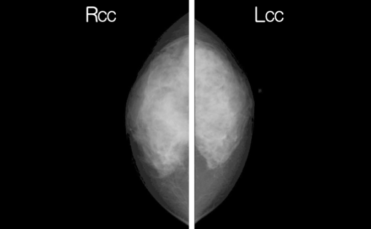
X-ray mammography, which is widely used as the global standard, is not applicable to women with high density breasts (a type of breast with high levels of collagen fibers that support the breast shape, which is 79% of Asian women under 50 years of age).
While capturing the images of high density breasts using X-ray mammography, the fibers are displayed as whitish lumps. Because breast cancer is also displayed in whitish form, its detection becomes extremely difficult. In fact, the detection rate is said to only be approximately 60%.
Although magnetic resonance imaging (MRI) scans and echocardiography can also be used in addition to X-ray mammography, they suffer issues in terms of contrast media and depth quantification.
Examination is extremely painful
X-ray mammography can be extremely painful, as it requires strong pressure to be applied to the breast during the examination, and this pain can serve as an obstacle for women undergoing breast cancer screenings.
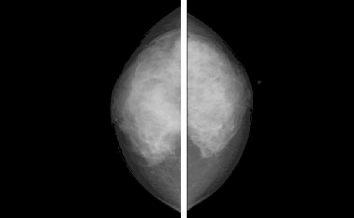
Results of measurement using conventional mammography
Risk of exposure
The risk of exposure exists because X-ray mammography uses radiation. Hence, this test is not available for pregnant women, or potentially pregnant ones.
Necessity of using a contrast agent
In MRI scans, one needs to use contrast agents that have a small contrast between abnormal and normal on the image and a high potential for side effects.
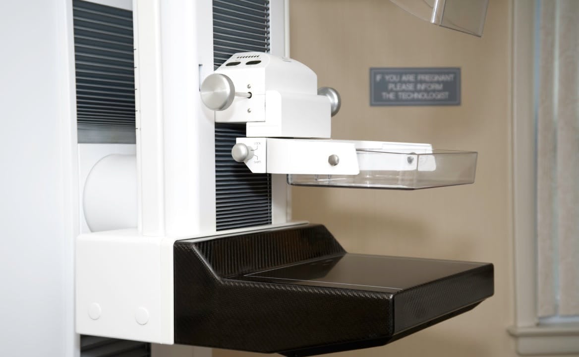
Measuring equipment for convention X-ray mammography
Developing novel solutions
IGS Approach to Issues
World’s first computational theory created by IGS to inversely analyze cancer via microwave waveforms
The microwave mammography developed by IGS uses the strong reflection of microwaves off cancerous tissue in breasts. Breast cancer has a higher amount of water molecules compared with normal tissues, resulting in higher relative permittivity. Microwaves emitted to the breast reflect at the border of breast cancer, where the relative permittivity is high, so it becomes possible to detect breast cancer by measuring the reflection.
Microwaves penetrate to the greatest depths in the breast and are often reflected by breast cancer. Conventional techniques have failed to determine where the microwaves hit the cancer cells and identify the true nature of the same. However, with Professor Kenjiro Kimura elucidating the previously unsolved “inverse problem of wave scattering” in applied mathematics, it has become possible to inversely analyze cancer using waveforms.

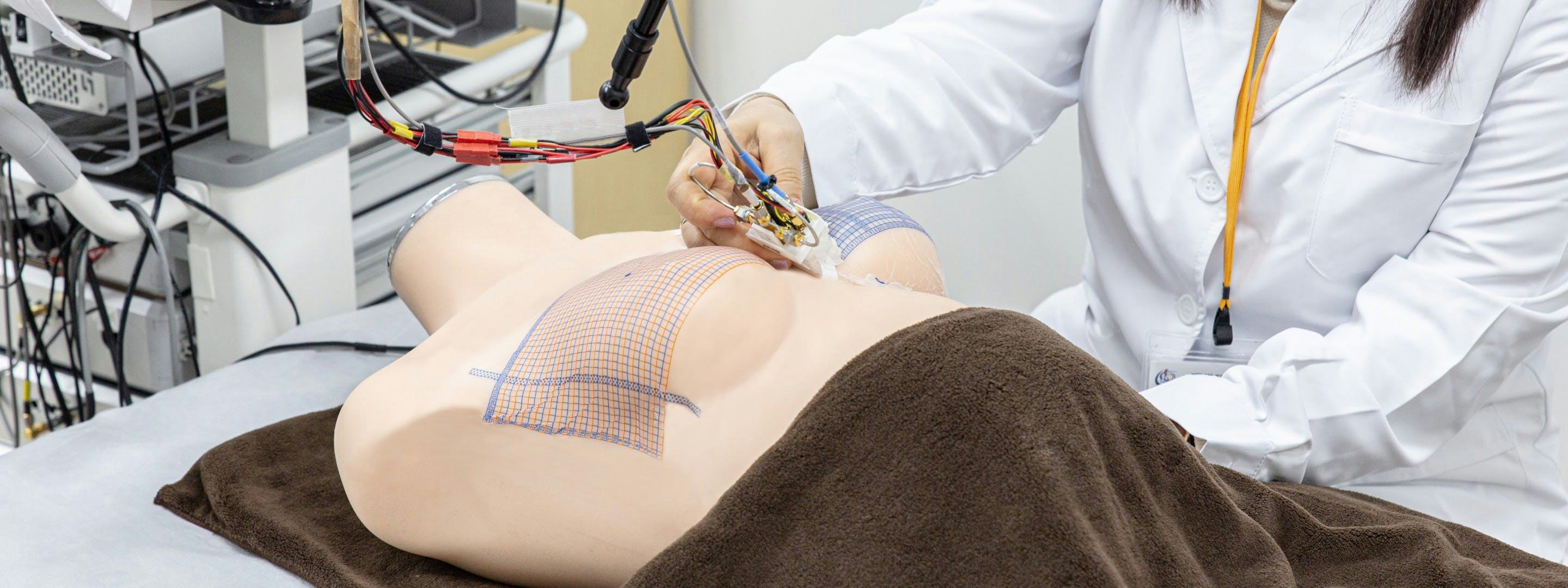
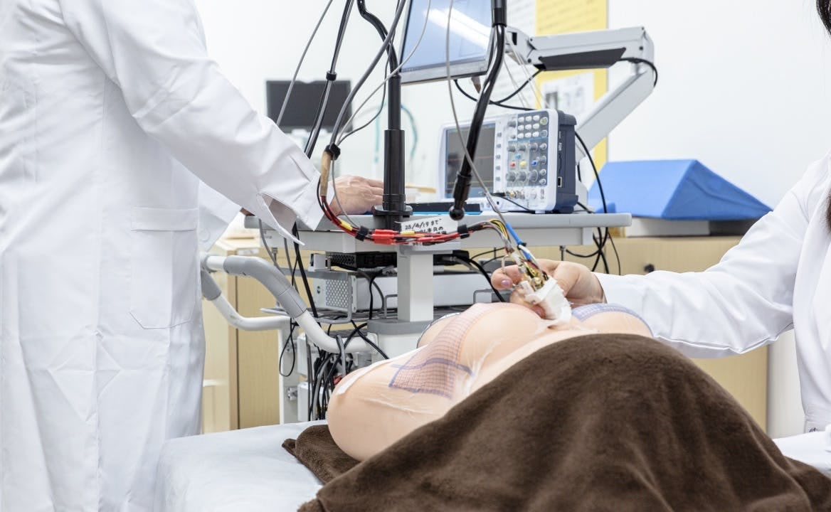
The examination is painless and takes approximately 10 to 15 minutes
Microwave mammography involves tracing a probe containing an antenna along the breast surface. This probe transmits and receives microwaves, scanning the entire breast area and outputting the results to a computer.
Unlike traditional mammography, this method does not require compressing the breast, ensuring a comfortable experience. The straightforward procedure of tracing the breast surface guarantees consistent imaging results, regardless of the operator, making the process highly convenient.
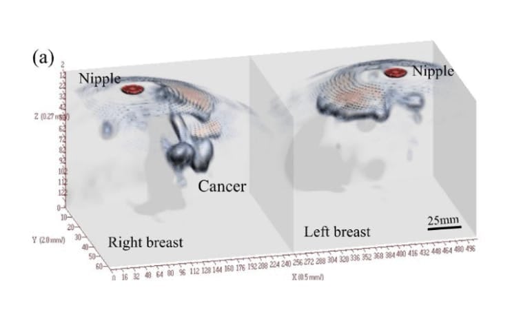
Precise positional identification using 3D images
Microwave mammography leverages scattered microwave data to generate 3D images, accurately displaying the position and size of cancerous tissue within the breast. This technique produces images with exceptionally high contrast, significantly enhancing the detection rate of breast cancer.
Advantages of IGS microwave mammography
IGS microwave mammography utilizes scattered microwave data to create 3D images, accurately pinpointing the location and size of cancerous tissue within the breast. The images produced have an exceptionally high contrast ratio, ensuring a high cancer detection rate. Additionally, this method does not require breast compression, making the examination painless and eliminating the need for radiation exposure. A clinical trial is currently underway at IGS to further evaluate the efficacy and safety of our microwave mammography technology.
Conventional X-ray mammography
Yes
Yes
2-D
IGS microwave mammography
None
None
3-D
Yes
None
Yes
None
2-D
3-D